About Title Color Change

Knee Pain

The knee, being the largest joint in the body, is a complex structure composed of bone, cartilage, and ligaments. The cartilage within the knee serves as both a cushion and a gliding surface. In a healthy state, this cartilage prevents the bones within the joint from rubbing against each other. Unfortunately, conditions like arthritis, injuries, aging, and degenerative issues can lead to the breakdown of cartilage. When arthritis affects the knee, bones come into direct contact, resulting in pain. For effective knee pain treatment in Vadodara, addressing these underlying issues is crucial.
About Title Color Change

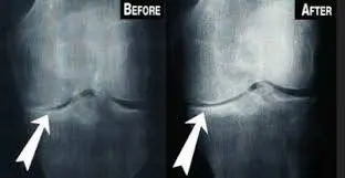
PRP Knee Injection Before And After


  • Prolotherapy and Prolozone Therapy: Injection of tissue proliferates (like ozone, dextrose, etc.) inside the joint and around the joint reduces pain, inflammation and strengthens ligaments. It is also claimed that they promote cartilage growth.
  • Visco-supplementation: High molecular weight hyaluronic acid resembling synovial fluid is very helpful particularly in early osteoarthritis with knee pain.
  • Intra-articular injections: Patients with severe pain of the knee, joint effusions, and local signs of inflammation benefit from intraarticular analgesics (pain killers). This will be effective for a short-term period in reducing pain and increasing quadriceps strength. Some patients will require about 2 to 3 injections in a year, using aseptic precautions, the infection rate is negligible. Sometimes mild flare-ups are possible in joint inflammation following intraarticular injections. Repeated injections are not recommended for the fear of damaging the cartilage of weight-bearing joints.
  • Radio-Frequency Procedure: This procedure is a very effective procedure in knee pain treatment and is approved by the US FDA. Genicular nerves carrying knee pain are blocked with this procedure. By blocking the nerve, the patient can feel pain relief for a long time. This type of treatment is useful for aged patients having severe arthritis knee pain and not able to go for surgery due to some high-risk disease.
Other Non-Interventional Therapies
  • Basic Electrotherapies like TENS, Ultrasound Therapy, IFT, Infrared therapy etc.
  • Advanced Electrotherapies like Low Level Laser Therapy, Radial Shock Wave Therapy, Pulsed Electromagnetic Field Therapy etc.
  • Dry Needling
  • Acupuncture
  • Non-Invasive Pulsed Radio Frequency Therapy
Surgery
  • When patients experience severe symptoms that are unresponsive to medical treatments and interventions, surgical options like tibial osteotomy, arthroplasty, and joint replacement may be considered. However, it is advisable to delay surgical interventions as much as possible, as total joint arthroplasty has a lifespan of approximately 10-20 years. Finding the best knee pain surgeon in Vadodara is crucial for informed decision-making and exploring suitable options. It’s important to note that patients undergoing surgical procedures may need to make lifestyle modifications due to the ergonomics of the replaced joint.
Title Color Change

Common Causes Of Knee Pain

Osteoarthritis: Osteoarthritis is commonest cause of knee pain. Up to 40 percent of the population may have knee osteoarthritis, or “wear and tear” arthritis. Osteoarthritis is one of the commonest joint problems with 80% of general population of radiologic evidence of osteoarthritis by 65 yrs of age. The disease process starts by age of around 20 yrs and manifest around 40-50 yrs. More than 40% of sufferers have no radiological evident of osteoarthritis.
Risk Factors:
  • Age
  • Female
  • Obesity (most important modifiable)
  • Previous knee injury
  • Lower extremity mal-alignment
  • Repetitive knee bending
  • Hereditary and nodal OA
  • Low level of vitamin C and D
  • Muscle weakness (quadriceps)
Pathogenesis :

The exact etiology is not known. By chemical stress on articular cartilage and subchondral bone leads to wear and tear to these structures. Thus, joint inflammation is very minimum compared to other arthritis and seen mostly in advanced disease. However, pain of osteoarthritis may be due to the following reasons:

  • Trabecular micro-fracture
  • Intraosseous hypertension
  • Periosteal irritation
  • Synovitis
  • Stretching of joint capsule and ligaments
  • Muscle spasm
  • Low-grade inflammation of the knee joint
Central sensitization contributes considerably to producing osteoarthritis of knee pain.

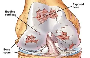
Pain around the joint that increases with weight bearing and movement and improves with rest is the commonest presenting symptom. It may be associated with morning stiffness and swelling of the joints. Clinical signs are tenderness, crepitus, joint effusion, decreased range of movement, valgus/varus deformity etc. X-ray shows decreased joint space, osteophytes formation (bone spur) and osteoporosis of subchondral bone.

Rheumatoid arthritis: Rheumatoid arthritis can affect joints on both sides of the body (both knees, both hands and/or both wrists). In rheumatoid arthritis, your body’s cells attack your own tissues. Rheumatoid arthritis affects three to five times more women than men and often presents between the ages of 20 and 50.

Over time, rheumatoid arthritis can cause cartilage to wear away, swelling in the synovium, and excess fluid in the knee. In later stages, bones can rub against each other.

Bursitis: Bursitis is the inflammation of any of the fluid-filled sacs (bursae) protecting the body’s joints. This is usually caused by repetitive motions or by a stress such as kneeling. Sometimes, a sudden injury can cause bursitis.

Tendonitis: The tendons – rope-like tissues connecting muscles to bone at the knee and other joints – can become painfully inflamed by repetitive and strenuous movement. Tendonitis is a common sports injury, caused by overuse of the same parts of the body. Patellar tendinitis, or “jumper’s knee,” is an inflammation or irritation of the tendon between the knee cap and the shin bone.

Patellofemoral pain syndrome (PFPS): Knee pain or discomfort while walking up and down stairs, jumping or squatting may be symptoms of patellofemoral pain syndrome. This common knee problem is felt toward the front of the knee. It can cause a grinding sensation when bending or straightening your leg, and can cause the knee to occasionally buckle. Sometimes called “runner’s knee,” patellofemoral pain syndrome may be caused by a kneecap that is not aligned properly, overuse, injury, excess weight or when the cartilage in the knee cap is worn significantly.

Injuries: Knee injuries can be the result of sports, falls or trauma. They typically involve the ligaments that hold two of the bones of the knee – the femur and tibia – together eg-ACL, MCL, & Meniscal injuries.

  • Diagnosis of Knee Pain
  • Careful medical history which includes:

    • A description of knee pain (aching, tenderness, burning or swelling)
    • Where the pain is located and when it occurs?
    • When did the pain start (and if it is the result of an injury or accident)?
    • Anything that makes the pain worse or better
    Investigations Conducted for Knee Pain

    X-rays, Sonography, MRI, CT Scan

    Blood Investigations – Depending upon the history and examination one can be prescribed for blood investigations like Uric Acid, Rheumatoid factor, Anti CCP antibody, Anti Nuclear Antibody, CBC, CRP, Vitamin B12, Vitamin D, Calcium, etc.

    Treatment

    I. Non-pharmacological therapy

    • Posture Correction – Avoid crossed leg sitting, sitting on the ground, squatting, frequent uses of stairs.
    • Thermotherapy – Apply ice or heat on knee. Ice for acute pain and swelling, Heat for chronic pain.
    • Reduction of obesity – loss of weight decreases load on the weight-bearing joints and thereby it retards the disease process. Losing 1 pound of body weight resulted in 4 pounds of pressure being removed from knees.
    • Knee pain exercises – It is very useful for patients with osteoarthritis of the knee. Strengthening of quadriceps muscle improves knee pain and function.
    • Patients are advised to use knee caps and walking sticks. Knee caps to be worn during working hours. Walking stick to be held in the opposite hand of the affected joint. Thus, it reduces the load on the joint and it is associated with decreased pain and improved function.
    • Deformity stabilization – use of proper shoes for varus or valgus deformity transfers the load to the other compartment and retards the disease process.
    • Education: Encourage patients to participate in self-management programmes and provide resources for social support and instruction on coping skills.
    2. Pharmacological therapy
    • Paracetamol / acetaminophen is used as the first line of therapy. The dosage recommended is from 1500 mg/day to a maximum of 4000 mg/day. Though paracetamol does not have anti-inflammatory properties, still it provides good pain relief and osteoarthritis also does not show a major inflammatory component.
    • Those who do not get adequate relief with oral paracetamol should take weak opioids like tramadol, codeine, or dextropropoxyphene along with Paracetamol. There are several combinations of Paracetamol with opioids that are used for a prolonged period without significant side effects.
    • Next line of therapy is NSAIDs. Ibuprofen 1200 mg to 2400 mg/day is the first line NSAID. If the relief is not adequate, paracetamol is added up to 4 g along with ibuprofen. These medicines are not used for a prolonged period.
    • Co-analgesics like Duloxetine is the most commonly used co-analgesics. This approved by US FDA for OA knee.
    • Nutraceutical supplements like Glucosamine, Chondroitin, MSM, Unnatured Type 2 collagen, Extracts of boswellia, turmeric, Rosehip etc. provide certain benefits over placebo though not studied extensively.
    • There are some so-called disease-modifying agents that had generated a lot of interest, claiming that some of them may help in the regeneration of cartilage and others inhibit degeneration. Others are diacerein, doxycycline, etc. Recent Studies do not recommend these groups of medicines and they do not help in regeneration.
    3. Interventions
    • Platelet Rich Plasma Injection – PRP therapy involves injecting platelets from the patient’s own blood to rebuild a damaged tendon or cartilage. It has been successful in not only relieving the pain, but also in jumpstarting the healing process. The patient’s blood is drawn and placed in a centrifuge for 15 minutes to separate out the platelets. The platelet-rich plasma is then injected into the damaged portion of the tendon or cartilage.